For referring doctors
Refer a patient.
This is the page we would want if we were referring. The form, exactly what imaging to send, what comes back to you, and a number that reaches a surgeon rather than a queue.
01 · Reach a surgeon
Four ways in, in the order we would use them.
If it is urgent, do not use the form. Call. The referral line is picked up by a surgeon or put through to one, and a ten-second conversation moves a patient into one of the assessment slots we hold open every week.
Urgent — the same week
Referral line
Suspicious mucosal lesions, spreading infection, dentoalveolar trauma, a patient you are not comfortable watching. We hold assessment slots for these every week and they are frequently unused.
Routine
02 · What to send with a case
Nine things, and one that matters more than the rest.
The clinical question. "Impacted 38" tells us what you can see. "Recurrent pericoronitis, third episode, patient is a flight attendant and wants it dealt with before the winter roster" tells us what to do about it.
- Patient name, date of birth, telephone number and preferred contact method
- Your practice name and the best number to reach you on, not the front desk
- What you want us to do — assess, treat, or give an opinion. "Please see and treat" is fine; "please see" is not always enough
- The clinical question you actually want answered
- Current radiographs, and the DICOM rather than a screenshot if you have a CBCT
- A full medication list including anticoagulants, antiplatelets and antiresorptives
- Relevant medical history: cardiac, respiratory, bleeding disorders, immunosuppression, pregnancy
- Whether the patient has an existing altered sensation — much easier to record now than to attribute later
- Whether the patient has a strong view about sedation
03 · Imaging requirements
What we need, by case type.
We would rather look at ten panoramics and say no to nine than not see the tenth. If you are unsure whether a film crosses the threshold for cross-sectional imaging, send it and ask — that is not an imposition, it is the job.
| Case | Minimum imaging | Also send | And tell us |
|---|---|---|---|
| Third molars | Panoramic under 12 months | CBCT DICOM if any canal marker is present | Pericoronitis episodes, trismus, sedation preference |
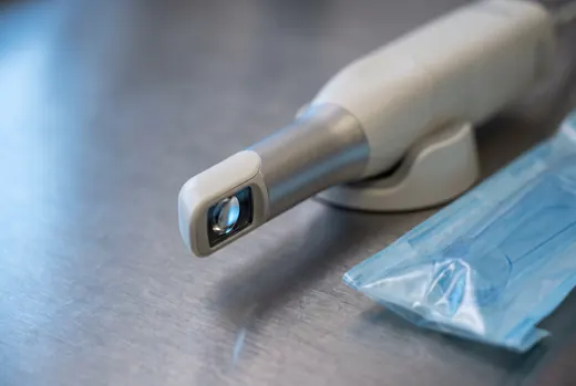
| Single implant | CBCT, small field of view | Intraoral scan or models, both arches | Restorative plan: screw or cement, who restores |
| Full arch | CBCT with the existing denture worn | Scan or impressions, both arches | Full-face photographs at rest and full smile |
| Grafting / sinus lift | CBCT showing residual height and septa | Photographs and probing chart for soft tissue | Sinus history: sinusitis, prior FESS, obstruction |
| Orthognathic | CBCT of the facial skeleton + lateral ceph | Scan of both arches, CR bite registration | The orthodontic plan and decompensation endpoint |
| TMJ | Panoramic as a screen; MRI for disc position | CBCT only for suspected osseous change | Pain history and what has already been tried |
| Pathology | Periapical or panoramic if there is bone involvement | Clinical photographs with a scale | Site in millimetres, duration, tobacco and alcohol use |
The five markers that mean CBCT
On a panoramic, any of these where a third molar root crosses the inferior alveolar canal:
- darkening of the root
- interruption of the cortical white line of the canal
- diversion of the canal
- narrowing of the canal
- narrowing or deflection of the root
Radiation, honestly
A cone beam scan carries a materially higher effective dose than a periapical or a panoramic, and a much lower one than a medical CT. We take a small field of view by default and we do not take one unless it will change what we do.
If you have already taken an adequate CBCT, send us the DICOM and we will not repeat it.
04 · The referral form
Send us a case.
Everything marked required is genuinely required; nothing else is padding. Attach imaging by replying to the acknowledgement email, or fax it to (612) 555-0151 with the patient's name on the cover sheet.
What comes back to you
Who you will be talking to
05 · Where we stand
Policies you will want to know before you refer.
None of this is controversial. It is written down because these are the questions that arrive on referral forms, and because a practice that will not answer them in public is harder to plan around.
06 · Clinical notes
Written for you, not for search engines.
Third molars
Five radiographic markers that mean we want a CBCT
A panoramic film answers most third-molar questions. These five findings are the ones that mean it has stopped answering them.
Dr. Ingrid H. Vestergaard · 6 min
Medical management
Anticoagulants: why we rarely ask patients to stop them
Interrupting anticoagulation to prevent a bleed that can be managed locally trades a small, controllable risk for a large, uncontrollable one.
Dr. Marcus O. Adeyemi · 7 min
Implants
Ridge preservation: refer before you extract, not after
The best time to decide about a graft is while the tooth is still in the mouth. Six months later, the decision has already been made for you.
Dr. Paul S. Lindqvist · 5 min