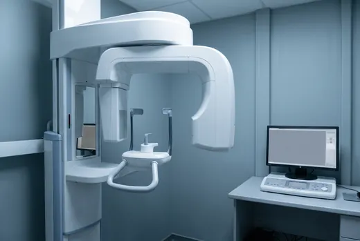
A panoramic radiograph is a two-dimensional projection of a three-dimensional relationship, and for most mandibular third molars that is entirely adequate. The value of the panoramic is not that it locates the inferior alveolar canal — it cannot — but that it tells you reliably when the canal and the root are not near each other. Where it stops being able to say that, it hands the question to cross-sectional imaging.
The five markers
These are the classical panoramic signs of a close relationship between the third molar root and the inferior alveolar canal. Any one of them changes what we want to see.
- Darkening of the root where it crosses the canal — the most frequently cited and, in most series, the most predictive of true contact.
- Interruption of the cortical white line of the canal, on one or both walls.
- Diversion of the canal from its expected course as it passes the tooth.
- Narrowing of the canal at the point of crossing.
- Narrowing or deflection of the root itself, or a dilacerated apex hooking around the canal.
None of these is a diagnosis of contact. They are a statement that the panoramic can no longer exclude it, and that the consent conversation needs better information than a projection can give.
What the CBCT changes
Cross-sectional imaging answers two questions the panoramic cannot: where the canal sits relative to the roots in the buccolingual plane, and whether there is intact cortex between them. A canal lying lingual to the roots, or running between them, changes the operative plan — and it is the finding that most often turns a planned extraction into a discussion about coronectomy.
| Finding | Typical plan |
|---|---|
| Canal separate, intact cortex | Standard removal. Consent for altered sensation as usual. |
| Canal in contact, cortex intact | Careful sectioning, no apical pressure. Consent emphasised. |
| Canal grooving the root | Sectioning and staged elevation. Coronectomy discussed. |
| Canal between the roots or perforating | Coronectomy is usually the safer offer where the tooth is vital. |
What we would like from you
- The panoramic itself, under twelve months old.
- The DICOM if you have already taken a CBCT — not a screenshot of the software. We want to reslice it.
- The clinical history: how many episodes of pericoronitis, whether there has been trismus, and whether the patient has an opinion about sedation.
- Whether the patient has any existing altered sensation. It is much harder to attribute afterwards than to record beforehand.
If you are not sure whether a film crosses the threshold, send it. We would far rather look at ten panoramics and say no to nine than not see the tenth.